Connecting To The Server To Fetch The WebPage Elements!!....
Anaesthetics act in quantum channels in brain microtubules to prevent consciousness
Travis Craddock, Stuart Hammerof, Jack Tuszynski et al, Nova Southeastern University, University of Alberta :: www.benthamscience.com/journal/
Recent research into the binding of anaesthetic molecules looks to revive the idea of hydrophobic pockets in the microtubules of neurons as the most suitable binding site for anaesthetic molecules. This could point to a latter day debunking of numerous but often superficial attacks on the proposal.
An early study showed that the strength of structurally different anaesthetics correlated with their solubility in a non-polar hydrophobic medium. Later in the 1980s, a study by Franks and Lieb showed anaesthetics acting within proteins. In particular they indicated that anaesthetics could also bind in lipid-like hydrophobic pockets within proteins. Post-synaptic receptors have been the favourite candidates for such proteins, but studies have not been conclusive in this respect. As a result, some opinion reverted to the hard to justify 19th century idea that all anaesthetics acted in different ways.
Molecular modelling has indicated pi-electron energy transfer through tryptophan. Further, anaesthetics molecules are modelled as binding to tryptophan channels. The energy transfer seen in tryptophan is similar to that seen in quantum coherent activity in photosynthesis. Anaesthetic molecules can impede pi-electron energy transfer and such molecules binding in this area may account for the selective action of anaesthetics on consciousness.
In microtubule-related consciousness theory, the binding of the anaesthetic molecules to a hydrophobic pocket by London forces disperses other dipoles that are involved in driving consciousness. This is called the quantum mobility theory of anaesthetic action. Research in physical chemistry is stated to show that anaesthetics bind via van der Waal London dipole forces involving the coupling of pi-electron clouds. Anaesthetics do bind to proteins within the lipid membrane of neurons, but research into this binding has not demonstrated this binding to have a significant effect.
Rod Eckenhoff's team has found anaesthetic action in the microtubules of neurons. The group surveyed all proteins that could bind with anaesthetics, and they found that anaesthetic molecules could bind with 23 membrane proteins and 34 cytoplasmic proteins. The latter included proteins in the cytoskeleton including actin and tubulin, the building-block protein of microtubules. Amongst these gene expression was particularly found to be altered in tubulin. While binding affinity is lower for tubulin than for membrane proteins, there are many more tubulins to act on, and this allows for significant occupation of tubulin binding sites by anaesthetic molecules.
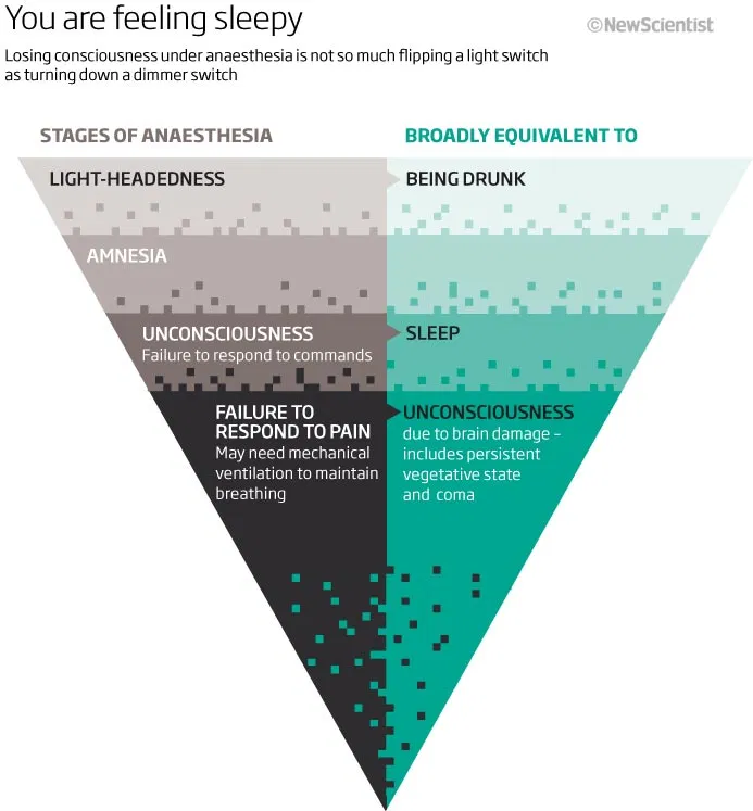
Amnesia is one effect of anaesthesia. The formation of memory is conventionally linked purely to changes in synapses, but a study by Janke and Kneussel suggested it could be encoded in the more stable microtubules of dendrites. Memory degeneration diseases such as Alzheimers are related to microtubule disintegration. Post-operative cognitive disfunction (POCD), due to repeated anaesthetic exposure, also involves microtubule instability. Anaesthetics act by means of intra-molecular forces without breaking covalent bonds. Although anaesthetics are seen as binding in non-polar hydrophobic pockets, they also possess polar groups capable of forming hydrogen bonds. Such polar groups could disrupt the configuration of proteins that depends on hydrogen bonds. This could be a factor in POCD.
If anaesthetics act in hydrophobic pockets in proteins this suggests that such pockets relate to a mechanism for supporting consciousness. London force bindings of anaesthetics within the microtubule proteins could hinder this pi-electron flow, and thus disrupt consciousness. It has become apparent over recent years that quantum superpositions or entanglement are functional in a number of different ways in organisms. Pi-resonance clouds in microtubules enable dipole oscillations, pi-electron mobility and an extended quantum wave function.
A map of pi-resonance clouds has been undertaken for tubulin by mapping the aromatic amino acids within tubulin. Each tubulin has a network of eight tryptophan, 36 tyrosine and 42 phenylalanine residues across the whole protein. The arrangement and dipole properties of these are similar to those in photosynthetic systems that are quantum coherent. This indicates that the microtubule tubulins are quite likely to support quantum coherence. Tryptophan in particular provides dipole couplings that could be disrupted by anaesthetic molecules. Distances between tryptophan and the other aromatics in the tubulin network are small enough to be effected by London forces. Such networks have so-called pi-stacking arrangements stabilised by such London forces, and this stabilisation is most apparent at the sort of separation distances seen in tubulin. The dispersion energy of the London forces comes from induced dipoles in the phenyl or indole rings of aromatic molecules. The force is based on two induced dipoles with a small spatial separation between them. The non-polar aromatic rings in tubulin are suggested to provide quantum mobility channels for electrons. Such pathways may be aligned to pathways in adjacent tubulins
It is finally proposed that the suggestions here may be capable of being tested by technologies such as laser scattering that have been employed successfully in studying similar photosynthetic systems.